Momentum Microscopy
The term momentum microscopy (MM) describes a modern approach to investigate the electronics structure of solids. MM experiments utilize a combined PEEM/ARPES spectrometer to couple lateral information of small sample sizes with the spectroscopic analysis of such, thus being a giant leap in the analysis of novel materials.
The basis of the instrumentation is a dedicated immersion lens being optimized for spectroscopic resolution at high lateral resolving power. An extraction field collects electrons with an emission angle of up to ±90°, thus collecting the complete photoemission signal for the sample. The immersion lens projects the sample information either in real space mode (PEEM) or in reciprocal space mode (ARPES and momentum microscopy). In general two projections are possible: a constant energy map showing a real space image or a constant energy surface of the samples band structure (MM), or an energy dispersive image of a real or reciprocal space coordinate (ARPES). The experimental data is then saved as a 3D dataset of the photoemission intensity, as a function of the kinetic energy and momentum/real space coordinate: IPES(EKin, kx/x, ky/y).
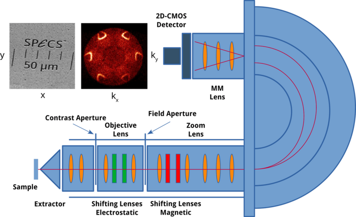
The big advantage of a momentum microscope over a classical hemispherical analyzer is the intrinsic coupling of the real space information with the spectroscopic data. Using field apertures inside the immersion lens, it is possible to collect photoemission data from a freely chosen small area of the sample, thus allowing ARPES investigations from smallest spots of down to 2 µm.
Momentum Microscopy performs well with advanced spin detection technologies. The MM lens technology is compatible with the direct imaging spin technology, for spin and momentum resolved mapping. The KREIOS 150 electron momentum spectrometer is compatible with the well-established 2D ARPES/3D Spin Detectors.
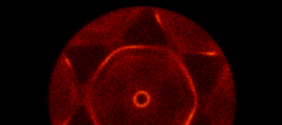
MM Mode
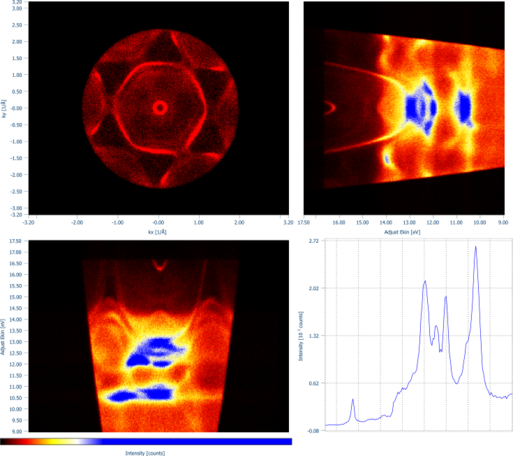
The MM mode displays a constant energy map on the detector in the dimention of reciprocal space. Thus angular emission is directly transformed into k-space. By scanning the energy, a full 3D dataset including the kinetic energy can be measured.
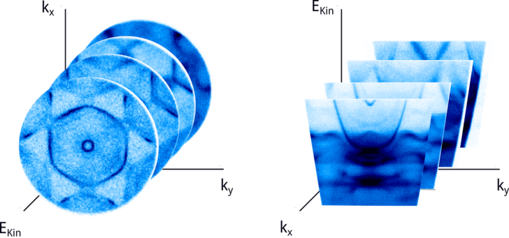
ARPES Mode
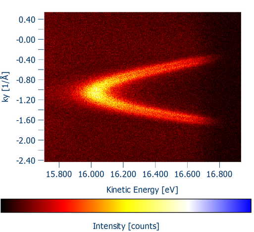
The advanced lens optics allow to project an energy dispervice image, e.g. a classical ARPES image on the detector. This is the standard operation mode for the KREIOS 150 but also available in the dual operation mode of the KREIOS 150 MM
PEEM Mode
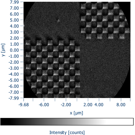
In PEEM operation, a precise image of teh surface can be aquired to identify areas of interest. Placing small apertures down to 2 µm, then allows to select those areas for µARPES and µMM investigations.